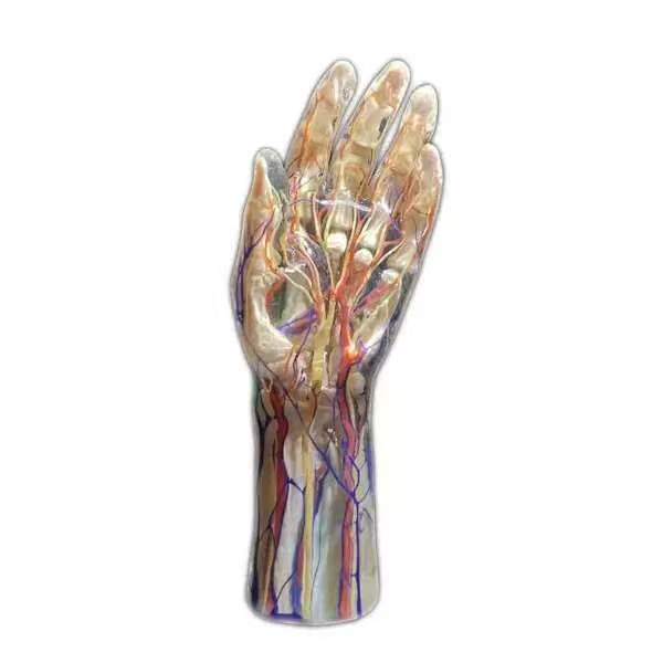
Medical institutions worldwide face a growing challenge in providing practical anatomical education. Traditional teaching relies heavily on human cadavers. However, physical specimens present serious hurdles regarding procurement, ethics, and high maintenance costs. Physical specimens are becoming scarcer and increasingly expensive to preserve.
Consequently, academic institutions are actively seeking innovative alternatives. These new educational tools must maintain clinical excellence without the accompanying liabilities. Digital anatomy systems successfully fill this void, offering medical students and professionals access to high-fidelity structures. This technology bridges the gap between theoretical textbook knowledge and clinical practice. Ultimately, it provides a safe, highly repeatable, and deeply immersive environment for studying the complex systems of the human body.
The Challenges of Conventional Specimen Training
For centuries, dissecting physical specimens has been the teaching standard, offering valuable tactile feedback. However, physical tissues present distinct limitations. Real tissue decays quickly, limiting student analysis of delicate structures. These structures include cranial nerves, deep vascular networks, and subtle lymphatic pathways. Furthermore, a physical cadaver represents only a single pathological state, age, and gender. This fails to show the vast anatomical variations found in actual clinical patients.
Preparing and maintaining wet labs requires specialized ventilation and preservation chemicals. It also demands strict biohazard disposal protocols. These ongoing expenses place a heavy administrative and financial burden on university training centers.
Advanced Interactive Features of a Modern Virtual Dissection Table
To address these educational and operational challenges, pioneering enterprises have designed sophisticated digital visualization systems. DIGIHUMAN is one such enterprise, dedicated to high-precision medical education. Integrating advanced 3D reconstruction algorithms with high-performance hardware creates an immersive educational platform.
This virtual dissection table provides students with repeatable opportunities to interact with the body. Users perform virtual dissections multiple times without deteriorating physical specimens. The system allows peeling away layers from skin to bone. With intuitive multi touch gesture controls, users can rotate and translate models. They can also zoom in on intricate systems with ease. This interactive approach helps students conceptualize spatial relationships.
Unmatched Scientific Accuracy and Engineering Specifications
Reconstructed from real human tomographic data, the system features ultra high precision cross sections with 0.1 mm accuracy. This detail is derived from continuous, high definition tomographic sequences. It represents over 17,571 layers for males and 16,141 layers for females. These layers clearly show the finest human structures. Such details may be difficult to observe consistently during traditional dissection.
The hardware features a high performance computer workstation to handle massive data files smoothly. This workstation includes an Intel Core i7 processor, 64GB DDR4 memory, a 2TB NVMe SSD, and an NVIDIA RTX 3080 graphics card. This setup supports smooth real-time rendering of 3D models. Depending on requirements, institutions can select different screen configurations. The ECDH Int II 88 model boasts a massive 88 inch infrared touch screen. This model offers a 3840×1080 resolution, a 1100:1 contrast ratio, and a 700 cd/m² brightness. The ECDH Int II 55 model features a 55 inch capacitive screen with a 4K resolution of 3840×2160. This variant supports up to forty simultaneous touch points for collaborative team learning.
Integrating Diagnostic Imaging and Clinical Case Modules
Modern medical training must connect anatomical knowledge with clinical diagnosis. The DIGIHUMAN platform includes over 180 clinical cases, integrating histories, descriptions, and over 1,700 CT or MRI images. Students can compare healthy 3D structures directly with pathological variations. This helps them develop critical diagnostic skills early in their training curriculum.
Furthermore, the system is DICOM compatible, allowing professors and radiologists to import actual patient data directly. It converts standard gray scale scans into interactive, three dimensional models. By bringing real world diagnostic imaging into the anatomy classroom, students learn to interpret scans in a clinical context. This prepares them for future residency programs and surgical planning.
A Versatile Ecosystem for Diverse Scientific Disciplines
The utility of this platform extends far beyond systematic human anatomy, offering seven core modules. These include gross anatomy, embryology, ultrasound, and animal anatomy. This makes the system valuable for agricultural and veterinary schools.
The platform is compatible with augmented and virtual reality devices, allowing users to experience anatomy in a holographic space. The slice library offers adjustable magnifications of histological specimens up to 40X. This feature gives students a cellular perspective on healthy and diseased tissues. Such versatility ensures that a single hardware investment supports multiple academic departments, maximizing overall utility and educational return.
Empowering the Next Generation of Medical Professionals
Transitioning to digital dissection technologies represents a major leap forward in modern anatomy education. By removing the financial and physical constraints of traditional cadaver labs, schools can focus entirely on delivering high quality education. The combination of ultra high definition human data, advanced multi touch hardware, and extensive clinical resources ensures that students are well prepared for the complexities of modern medicine. As global educational standards evolve, integrating these advanced systems is key to training highly skilled, competent healthcare professionals.
